
Osteochondritis Dissecans (OCD)
- Diagnosis
- Non-operative Options
- Operative Options
- Before Your Surgery
- After Your Surgery
- Your Rehab
What is Osteochondritis Dissecans?
Osteochondritis dissecans is a joint condition in which a piece of cartilage, along with a thin layer of the bone separates from the end of the bone because of inadequate blood supply. The separated fragments are sometimes called “joint mice”. These fragments may be localized or may detach and fall into the joint space causing pain and joint instability.
Osteochondritis dissecans can occur in any of the joints including your elbows, ankles, shoulders and hips.
Osteochondritis dissecans is more common among boys and young men between 10 and 20 years who actively take part in sports. Athletes participating in sports such as gymnastics and baseball may develop osteochondritis dissecans.
What are the Causes of Osteochondritis Dissecans?
The exact cause for osteochondritis dissecans remains unknown and certain factors such as trauma, fractures, sprains, or injury to the joint are considered to increase the risk of developing the condition. Osteochondritis dissecans may be caused by restricted blood supply to the end of the affected bone that usually occurs in conjunction with repetitive trauma. Following the injury or trauma, the bones in the area may be deprived of blood flow leading to necrosis and finally the bone fragment may break off. This may initiate the healing process, however, by this time, articular cartilage will be compressed, flattened, and a subchondral cyst will be developed. All these changes in addition to increased joint pressure cause failure of healing of the joint.
Symptoms of Osteochondritis Dissecans
Patients with osteochondritis dissecans usually have elbow joint pain, swelling, stiffness, and decreased range of motion. Pain usually increases after activity.
Diagnosis of Osteochondritis Dissecans
Your doctor will probably order an X-ray of both the right and left elbow to see the abnormality in the joint space and to compare them. You may also have a CT or MRI scan that is useful in determining the location of loose fragments within the joint.
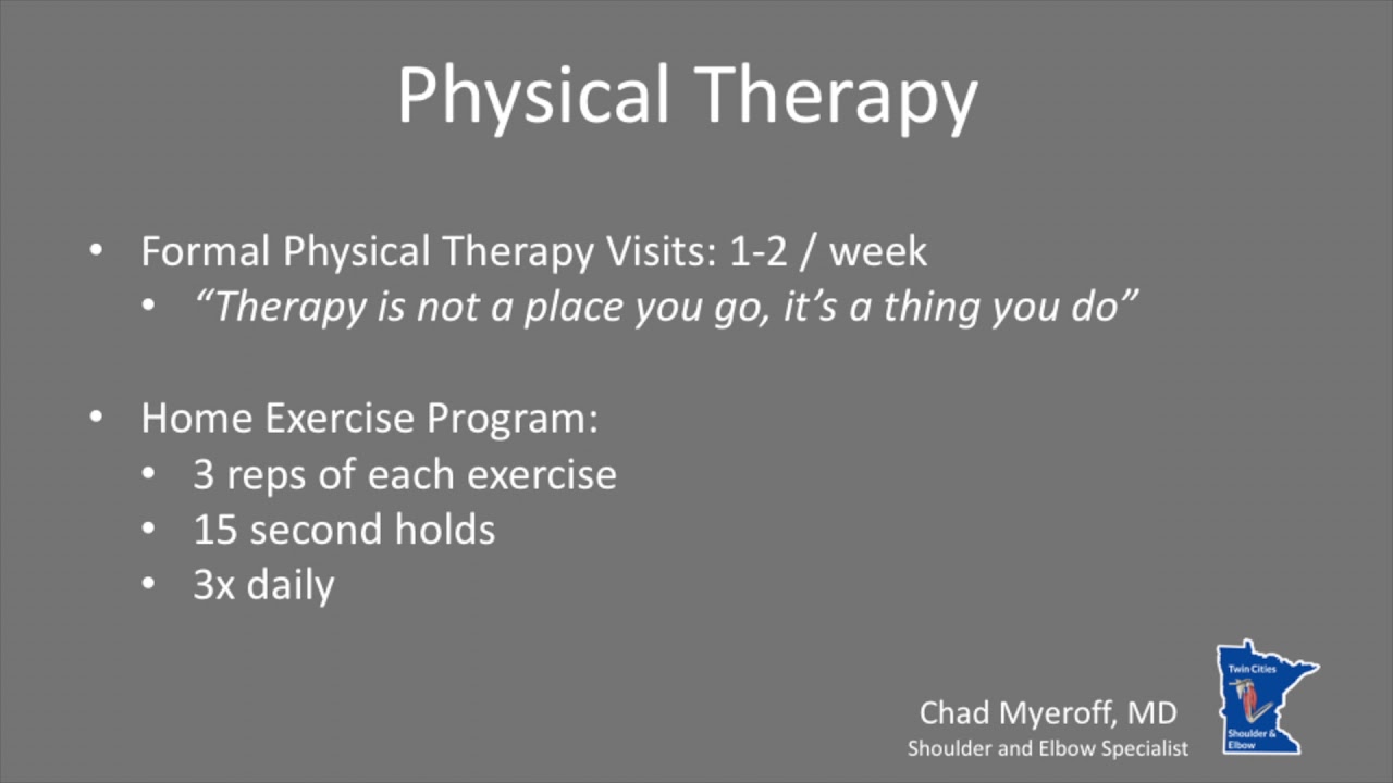
- Physical Therapy Intro
- Shoulder and Elbow Steroid Injection
Your physician may recommend various treatments depending on the reports of diagnostic scans, age, severity, stability of the cartilage and other factors. Goals of treatment are to relieve the symptoms and stop or impede the progression of degeneration of the joint.
Conservative treatment approaches such as wait & watch approach, pain medications, and immobilization for 1-2 weeks are recommended if the condition is diagnosed at an early stage and if the severity is mild. However, surgery is required if the condition is diagnosed at an advanced stage or if the condition is severe.
Want to know more?
Surgical correction of osteochondritis dissecans can be done using the open technique or arthroscopic technique. Some of the surgical procedures include drilling, bone grafting, open reduction internal fixation, osteochondral grafting, or autologous chondrocyte implantation (ACI).
- Drilling: In this method, multiple small holes are drilled into the bone which allows the growth of new blood vessels in the defective area. This promotes blood flow into the defective area, thereby initiating the healing response and formation of new cartilage cells inside the lesion.
- Open reduction internal fixation: Open surgery is performed in cases where the defective area is difficult to reach with an arthroscope. Hence, an open incision may be required. In this procedure, an incision is made in front of the joint to allow the surgeon to see the joint and the loose bodies are removed. Internal fixation involves fixing the fragments using internal fixators such as metal screws, pins, or wires.
- Bone grafting: It helps to fill the gap after removal of the dead or necrotic bone. In this procedure, bone graft is placed on the damaged site. This procedure may be performed to repair the damaged area or replace the missing bone. Autograft (harvested from the same individual) or allograft (taken from bone bank) may be required to help in the growth of a new bone.
- Osteochondral grafting: The procedure involves transfer of healthy cartilage plugs from the non-weight-bearing areas of the joint and transferring into the damaged areas of the joint in mosaic pattern. It allows the newly implanted bone and cartilage to grow in the defective area. Grafts may be taken from the same individual (autograft) or from a donor or bone bank (allograft).
- Autologous chondrocyte implantation (ACI): In this procedure, healthy cartilage cells are harvested from the non-weight-bearing joint of the patient and cultured in a laboratory. The cultured cartilage tissue patch will be implanted into the defective area which also promotes the growth of new cartilage.
Once you and your doctor decide that surgery will help you, you will need to learn what to expect from the surgery and how to actively participate in the treatment plan for the best results afterward.
Preparing mentally and physically for surgery is an important step toward a successful result. Understanding the process, and your role in it, will help you recover more quickly and have fewer problems.
Before surgery, your doctor will perform a complete physical examination to make sure you don’t have any conditions that could interfere with the surgery or the outcomes.
- Routine tests, such as blood tests and X-rays may be performed.
- Discuss any medications you are taking with your doctor as you may have to stop or alter your intake before surgery. If you are taking aspirin or anti-inflammatory medications or any drugs that increase the risk of bleeding, you will need to stop taking them one week before surgery to minimize bleeding.
- Discuss with your doctor about preparing for potential blood replacement, medical interventions and other treatments prior to surgery.
- Report any infections to your surgeon. Surgery cannot be performed until all infections have cleared up.
- If you smoke, you should stop or cut down as smoking interferes with wound healing and can affect your recovery.
- Have someone available to take you home, as driving is not recommended for at least 24 hours or as advised.
- You may need help with everyday tasks such as cooking, shopping and laundry.
- Put items that you use often within easy reach, so you won’t have to stretch and bend as often.
- After Surgery Video
- Elbow Surgery Recovery Video
- Elbow Elevation Technique Video
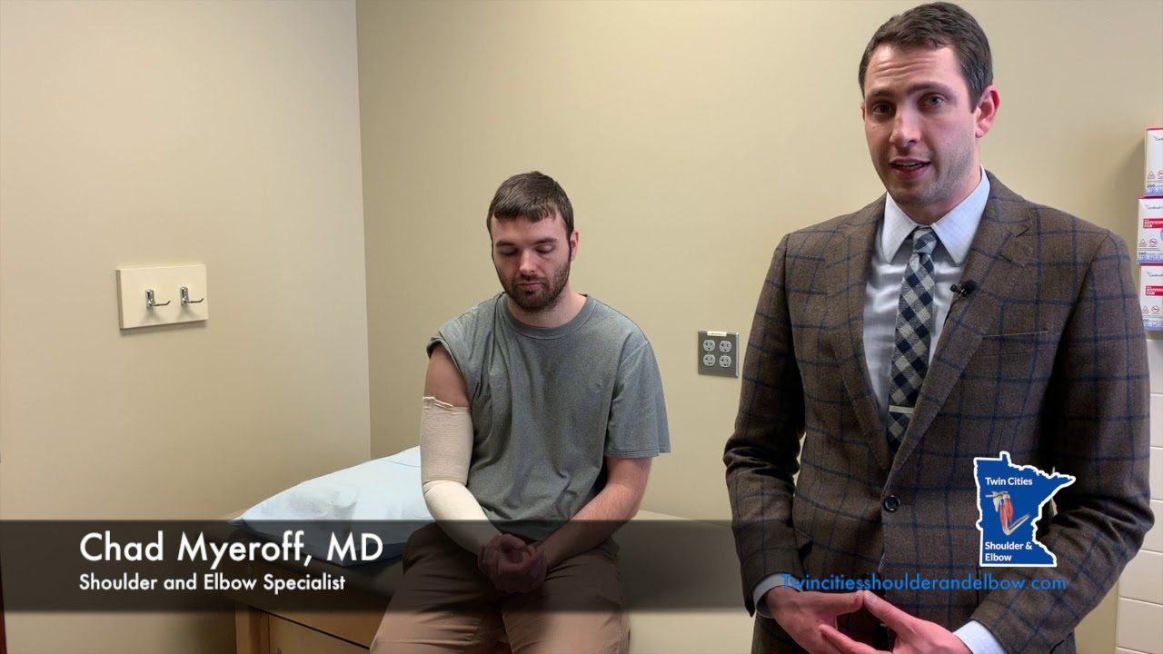
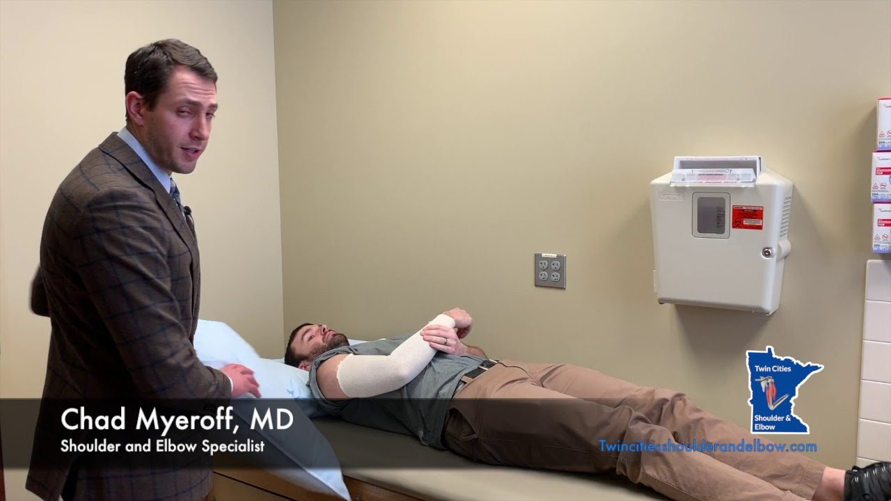
The post-surgical instructions include:
- Make sure to get adequate rest.
- Raise your elbow on pillows above the level of the heart to help reduce swelling.
- Keep the incision area clean and dry.
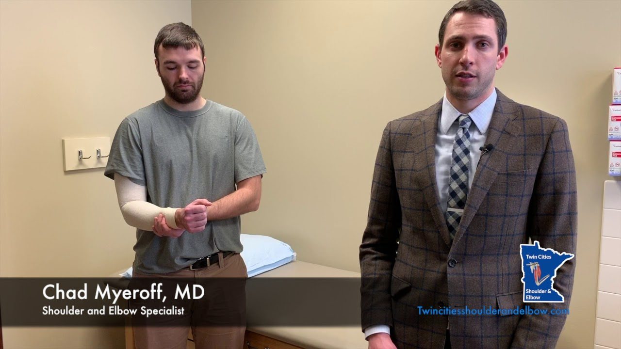
- A compressive stocking may be applied from the armpit to the hand once the dressing is removed to decrease pain and increase range of motion.
- Your doctor will prescribe pain medications to keep you comfortable.
- Eating a healthy diet and not smoking will promote healing.
Want to know more?
- Physical Therapy Intro Video
- Finger ROM Video
- Standard Elbow ROM Video